Locations Near You
Contact Us Today
Advanced Treatment Technologies
Advanced Linear Accelerators
State of the art Linear Accelerators deliver precisely doses of life-saving radiation while allowing doctors to choose the most appropriate form of treatment for each patient. Our Linear Accelerator equipment all feature VMAT technology for a quick and efficient treatment, We were the second radiation therapy center in the U.S. offering the revolutionary VMAT treatment at our facility in Plano, Texas. All Landmark Cancer Centers utilize state of the art Linear Accelerators from Elekta and Varian to provide you with the finest radiation therapy available today.VMAT Treatment
During a VMAT radiotherapy treatment, the treatment beam is continually shaped by a multi-leaf collimator (MLC), a device with 120 computer-controlled mechanical “leaves” or “fingers” that move to create apertures of different shapes and sizes so that the treatment beam conforms to the shape of the targeted tumor. A VMAT radiotherapy treatment is delivered very quickly, typically in less than two minutes, with just one turn of the machine around the patient. VMAT shapes and modulates a highly focused treatment beam so that it focuses on the tumor, sparing surrounding healthy tissue. It treats the entire tumor with pinpoint accuracy and is easier on the patient, who does not have to hold still for long periods of time.Intensity Modulated Radiation Therapy (IMRT)
In intensity modulated radiation therapy (IMRT), very small beams are aimed at a tumor from many angles. During treatment, the radiation intensity of each beam is controlled, and the beam shape changes hundreds of times during each treatment. As a result, the radiation dose bends around important healthy tissues in a way that is impossible with other techniques. Because of the complexity of these motions, Verity Radiation uses special high-speed computers, treatment-planning software, diagnostic imaging and patient-positioning devices to plan treatments and control the radiation dose during therapy. For IMRT to be effective, the anatomical position of the tumor and surrounding healthy tissues must be accurately defined. CT, PET scans and MRIs provide the necessary three-dimensional anatomical information, while advanced imaging devices provide daily information about the location of internal organs. A device called a multileaf collimator adjusts the size and shape of the computer-determined radiation beams. The collimator, a computer-controlled mechanical device, consists of dozens of individually adjusted metal leaves. These leaves move across the irradiated tissue while the beam is on, blocking out some areas and filtering others to vary the beam intensity and precisely distribute the radiation dosage.Image-Guided Radiation Therapy (IGRT)
Tumors can move, both during a radiation treatment session and from one treatment session to another. This commonly occurs as a result of normal internal organ action (digestion, elimination, and breathing). If these changes in position move the tumor out of the planned treatment range, the tumor may either not receive the full amount of radiation that it should, or normal tissues may receive more radiation than they can tolerate. By use of new image-guided techniques (IGRT), it is now possible to verify tumor locations each day. One of the most effective methods, used by Verity Radiation daily, involves producing a computed tomography (CT) image of the patient using the “cone beam” technique. By using a linear accelerator’s larger conical beam, the entire 3-D volume of the treatment area can be imaged with just one rotation of the device. Image-guided Radiation Therapy is an important key in achieving both unparalleled tumor control and normal tissue sparing. At Verity, we have the integrated tools that work toward successful image-guided motion management in the radiation oncology process to provide efficient and effective treatment.3D Conformal Radiotherapy
Each patient’s CT scan is imported directly into the treatment planning computer and the physician uses this 3-dimensional information to define the treatment area. In some cases, MRI and PET scans are merged with the CT to help define the tumor. The dosimetrist and physicist work with your physician to design radiation beams that conform to the shape of the tumor and avoid healthy tissue to the greatest extent possible. This allows us to deliver high doses of radiation to the tumor while minimizing the radiation to nearby normal tissues.Stereotactic Radiosurgery (SRS)
One of the major features available through use of the Trilogy system is Stereotactic Radiosurgery (SRS). In SRS, a number of precisely directed beams of ionizing radiation are aimed from diverse directions and meet at a specific point within the body, delivering very high doses of radiation to that point. As with other treatment techniques available at Verity, a CT scan is used to provide a 3-dimensional view of the tumor. This data is used to develop complex plans designed to deliver highly focused radiation while sparing normal adjacent tissues. In some cases an entire course of treatment is given in a single treatment.Advanced Imaging
Diagnostic imaging is the process of producing detailed pictures of organs and various body structures. These pictures are used to detect various abnormalities such as tumors, to determine the extent of a disease, and to evaluate the effectiveness of a treatment. Diagnostic imaging may sometimes be used when performing biopsies or other surgical procedures. All Landmark Cancer Center imaging departments are American College of Radiology accredited and staffed by trained, certified professionals. Diagnostic imaging capabilities vary slightly by Landmark Cancer Center location, feel free to contact us and inquire about our imaging capabilities by location.Advanced Chemotherapy
The primary tool of any Medical Oncologist is the use of either a single antineoplastic drug or the combination of such drugs into a standardized treatment regimen that kills cancer cells by stopping them from growing and dividing. Most chemotherapy is delivered intravenously, though some can be delivered orally. Our Oncologists determine how often you'll receive chemotherapy treatments based on what drugs you'll receive, the characteristics of your cancer and how well your body recovers after each treatment. Chemotherapy treatment schedules vary. Chemotherapy treatment can be continuous or it may alternate between periods of treatment and periods of rest to let you recover. The total number of treatments you receive depends on numerous factors. Chemotherapy treatments are given as outpatient treatments and involve a relatively small amount of time each day of treatment. Consultation visits may last for as much as an hour or more. It is important that you not miss treatments as this could affect treatment results. Chemotherapies work by killing cells that divide rapidly. As they kill fast-growing cancer cells, though, they can also damage or kill healthy cells as well. Damage to blood cells, for example, leads to side effects such as anemia, fatigue, and infections. Chemotherapy can also damage the cells that line the mucous membranes found throughout the body, including those inside the mouth, throat, and stomach. This leads to mouth sores, diarrhea, or other problems with the digestive system. And damage to cells at hair roots, or hair follicles, leads to hair loss. Each person with cancer reacts differently to chemotherapy and its various side effects. Fortunately, Landmark doctors now have many ways to reduce and even prevent these side effects. We will help with managing side effects so that your treatment goes as smoothly as possible.Elekta Infinity™ Treatment
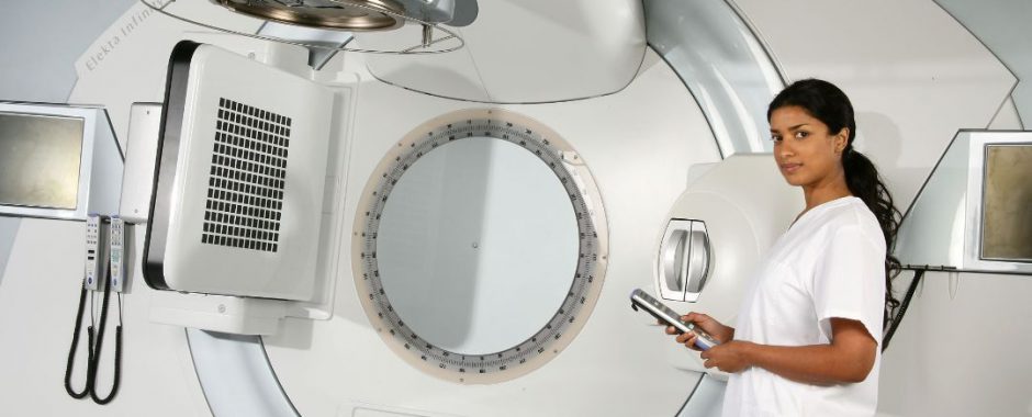
Comprehensive image-guided radiation therapy system with VMAT Elekta integrates hallmark Elekta Synergy® image-guided workflow in the advanced Elekta Infinity system, a comprehensive treatment system that includes Volumetric Modulated Arc Therapy (VMAT). Combining superior dose conformance and treatment speed, VMAT enables clinicians to “shrink wrap” the dose around a tumor by simultaneously manipulating the gantry position and speed, MLC leaves, dose rate and even collimator angle. 3-D image guidance complements VMAT’s speed (< 2 min. to 5 min.) by providing the clinical confidence to visualize targets with the patient in the treatment position minutes before therapy. Integrated imaging enables clinicians, on a daily basis, to correct patient setup errors due to organ motion and increase treatment accuracy. Imaging includes a variety of 2D, 3D and 4D X-Ray Volume Imaging (XVI) tools. Infinity offers a choice between single or multiple arcs – typically depending on the treatment site – to optimize the VMAT plan. Infinity with VMAT requires much fewer MU’s than with conventional methods, reducing total MU delivery by up to 50 percent. With its new MLC, Infinity optimizes VMAT delivery by limiting interleaf and overall patient leakage for lower dose volumes to critical structures.Varian Clinac iX™ Treatment
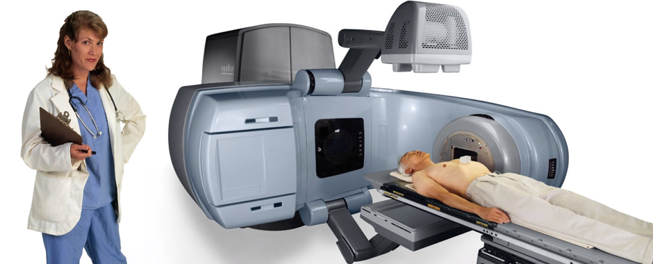
The Varian Clinic iX linear accelerator is a streamlined, high-performance, state-of-the-art platform that produces radiation upon demand for the treatment of cancers. It can produce up to 7 beams (two photon beams and five electron beams) for treating deep seated tumors. It offers a range of electron energies used to treat cancers closer to the skin, including the Head, Neck, Breasts, Skin, and other more superficial cancers. Furthermore, it allows for precise treatment of cancers, while limiting radiation dose to other organs and tissues by means of Cone-beam CT based imaging and repositioning, Multi-leaf collimation (MLC blocking) systems, and Intensity modulated radiation therapy (IMRT). This combination of quality and customization allows Landmark clinicians to meet the specific needs of each site.
Breast Cancer Survivors Story
Prostate Cancer Survivors Story
Contact Us
Locate The Center Nearest You
Landmark Cancer Centers
All Locations Open Weekdays 8:00a - 5:00p
Texas :: Oklahoma :: Arkansas :: Missouri
CONTACT US NOW
Always Dial 911 For True Medical Emergencies
Landmark Cancer Centers
All Locations Open Weekdays 8:00a - 5:00p
Texas :: Oklahoma :: Arkansas :: Missouri
CONTACT US NOW
Always Dial 911 For True Medical Emergencies
Testimonials
I personally was not pleased with my first visit. I was in pain and looking for sympathy and was confronted with cold facts about my condition which Dr. Gilbert was correct in doing. I am now completely satisfied with the treatment and would be happy to recommend Dr. Gilbert to anyone. He and the staff are just plain great.
, Plano, Texas
Two very special young people. They made my radiation visits very pleasant. Beverly and Denise also made my (33) visits great. Dr G was very nice also!
, Carrollton, Texas
Dr. Gilbert and the staff were great in listening to my treatment concerns. Both doctor and staff were very pleasant and took a lot of time with me. I really appreciate it and would like to thank everyone. Treatment progresses well and I know they went out of their way to make sure my treatment is the best available to me. Thanks!
, Addison, Texas
As an old army colonel I used to work with complex individuals and personalities, what I witnessed during the duration of the treatment was the perfect example of true teamwork and dedication. Everyone seemed to be enjoying what he or she was doing and eager to jump in at anytime, with a central objective in mind – to make the patient feel loved by people who really care. Please keep up your outstanding work!
, Plano, Texas
My treatment was good! Not much downtime and everyone was very nice! The Doctor was very knowledgeable and comforting. I feel very lucky that he happened to be the one there for my treatment.
, Lake Dallas, Texas
Who would ever say they looked forward to being treated for cancer??? Well I would! The entire staff went out of their way to make me comfortable – 42 times! Y’all are super – I will actually miss seeing you each afternoon!
, Lewisville, Texas
Dr. Gilbert- Thank you for taking care of me in my hours and days of need. You were very caring from the very start when I first met you during my initial consultation… Thank you for taking the extra time and effort to make sure I got the treatment I needed .
, Allen, Texas
Its hard to choose one particular person among the staff of angels at Verity. I thank God for Dr. Gilbert and the staff. Continue to do the wonderful job!
, Plano, Texas
The Doc is a very caring and nice person besides being an excellent doctor. He is very prompt, courteous and takes time with you. His work is excellent. I will recommend him to anyone. It is rare to find a doctor now-a-days who seems to really care about you.
, Frisco, Texas
Dr. Gilbert- You are now one of our favorite doctors, thanks so much for your tender respect during my wife's treatment.
, Carrollton, Texas
Dr. Gilbert - Our confidence in your knowledge and expertise, our confidence in YOU, got us both through a very difficult and frightening time. We thank our lucky stars every day to have found you!
, Farmers Branch, Texas
Shana and Vince are very courteous and caring. Denise went the extra mile in dealing with approvals for my prescriptions and submitting them to the local pharmacy. Dr. Gilbert was comforting while I was undergoing treatment because of his own personal experience.
, Flower Mound, Texas
Just a note to say how much I appreciate what you have done for me. I’m sure once this is all over, I will be in tip-top shape. For the past eight and a half weeks, I can’t believe how patient you were with me, even when I didn’t get on the table straight. You both have turned out to be good friends. I’m not sure what I’ll do in the mornings starting tomorrow. I sure wish the best for the both of you. Take care
, Little Elm, Texas
Patient Forms
As a first time patient we have made it easy for you by offering health related forms online. A single file download contains all of the appropriate form(s) required. Simply print them out and bring them to your first appointment already completed. By offering you this option to print the forms in advance, you will be able to complete them at your own pace and in the comfort of your own home. Please print the forms on white paper only.


